Episode 3: Anatomy and Pharmacology: Negotiating with the Greeks
A weblog entry by Shahrzad Irannejad, Jonny Russel, Aleksandar Milenković, Rebekka Pabst and Oxana Polozhentseva.
Hegemony of Greek Medicine and Pharmacology
So we landed in the land of knowledge. Where all “science” had once begun. But had it? We have recently been having discussions at our GRK about what constitutes science. Or “when” things “first” began. Or whether or not such questions yield valuable knowledge. Being physically in the land of the supposed “Greek Miracle” gave us the excuse to take a closer look at two contemporary scientific disciplines. Based on our common interests, Anatomy and Pharmacology were points of focus.
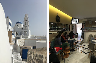
We used one long afternoon in Santorini to sit together and discuss various ways of conceptualization of the body in different cultures and different eras: from Ancient Egyptian looks into the body, over to the Ancient Greeks, the interlude in Alexandria, moving on to Medieval Europe to arrive at today.
 |
Fig 1.
The Inside and the Outside of where we sat down for our meeting in Santorini
(photos by Shahrzad Irannejad)
|
Apart from the Pharmacology Museum of the University of Athens, we had also planned to visit the Anatomy Museum; this plan was in fact our main incentive for putting together an “anatomy panel”. However, unfortunately, we did not manage to visit the Anatomy Museum. We were lucky enough that the Pharmacology Museum was open, despite the Easter holidays. Their hospitality at the Pharmacology Museum compensated for our bad luck and missing the Anatomy Museum.
Looking inside the Body
“Egyptian Conceptions of Internal Anatomy”, Jonny Russell
For the first look at the concepts of internal anatomy and dissection, we turned to the Ebers Papyrus (dated c. 1550 BCE). The text outlines a belief in conduits (often translated as ‘vessels’ or ‘Gefäße’) which — centred and controlled by the heart — were believed responsible for the flow of bodily fluids, abstract matter, and conceptualized substances believed responsible for pain and illness. Ancient Egyptian conceptualisations of the human body involved elements of biophysical observation, but these were nonetheless limited and do not support evidence for the practice of dissection or “scientific” examinations. Rather, the healing tradition appears to have operated on a phenomenological and culturally influenced theoretical basis.
"Pre-Alexandrian Dissection: A Myth?", Aleksandar Milenković
Although human dissection was a taboo in ancient Greece — before it was practiced in Alexandria — there is a peculiar report that suggests otherwise. Namely, in a commentary to Plato’s Timaeus dated around 400 AD, Chalcidius writes about the nature of the eye, where he states that Alcmaeon of Croton, a 5th century BC thinker, was the first to dissect the human eye. This report inspired many scholars to adorn Alcmaeon with titles such as the father of anatomy, physiology or even medicine. The lack of earlier sources that indicate the same shows us that Alcmaeon most likely did not dissect the eye, and his own theory that the eye contains fire and water, and that the eye is connected to the brain with certain passages, only implies that he conducted non-invasive experiments which do not require cutting out the eyeball.
 |
| Fig 3. Medieval manuscript of Calcidius' Latin translation and a commentary of Plato's Timaeus. Vatican, Reg. lat. 1308 fols. 21 verso - 22 recto. |
“Early Studies of Human Anatomy in Alexandria”, Rebekka Pabst
With the beginning of the reign of Ptolemy I (about 367-283 BC), Alexandria became the center of science in antiquity. Since Ptolemy I allowed dissections on human corpses for anatomical studies, a school of anatomy was founded in Alexandria, Egypt. Among the leading scientists of this school were Herophilos of Chalcedon (325-255 BC), Erasistratos (305-250 BC) and Eudemos of Alexandria (exact dates unknown). Scientific autopsies provided insights into human anatomy. For example, Herophilos described the structure of the brain and the eyeball. He also distinguished between arteries and veins. Erasistratos studied the cerebrum and cerebellum more intensively and finally realized that all nerves ultimately emanate from the brain. Eudemos, on the other hand, focused on the pancreas and also named individual human bones. However, the original records of these anatomists were completely destroyed during the first (48 BC) or second (391 AD) conflagration of the Library of Alexandria. Today, they are only partially preserved in the writings of later scholars, especially in the works of Galen (circa 130-215 AD).
 |
Fig 4. Athens and Alexandria: the two Scientific Centers of Antiquity. (image: Manley, Bill, The Penguin Historical Atlas of Ancient Egypt, Harmondsworth 1996, 129) |
"The Beginning of Dissection in the Middle Ages", Oxana Polozhentseva
The dissection of human cadavers for medical purposes experienced various cycles of legalization and prohibition in different countries of Medieval Christian Europe. The aim of the presentation was to show the complexity of beliefs and prejudices about post-mortem examination, that were circulating at this period of history. Dissection was rare during the Middle Ages, but it was still practiced: a few surgical texts, pictures, and conserved evidence of human dissections have survived. Different factors influenced the development in this area of knowledge, e.g. opinions on the subject officially expressed by the church, establishing of the European Universities (with Anatomy as one of the studied subject), development of forensic procedures and so on. The “leading” country in human dissection was Italy with its famous Medical Schools at Salerno, Bologna, and Montpellier. The first public dissection north of the Alps was held in Vienna in 1404.
 |
| Fig 5: Dissection scene, from ‘Le Livre des Proprietes des Choses by Barthelemy l’Anglais’, 15th century. Source |
“Anatomy as Spectacle”, Shahrzad Irannejad
Until the mid-1800s, anatomical study was the object of widespread reproach. It is only in the late eighteenth century that practical anatomy first began to displace the then-dominant tradition of speculative Galenic anatomy. From the late eighteenth century until the turn of the twentieth, exhibitions featuring anatomical displays of human bodies were consistently popular. To contextualize one such exhibition: anatomy museums attached to medical schools, Shahrzad drew on Elizabeth Stephens’s book entitled “Anatomy as Spectacle: Public Exhibitions of the Body from 1700 to the Present” (Liverpool University Press, 2011). From the perspective of cultural studies, the book examines displays of the human body from the anatomical Venuses of the 18th century, trough popular museums, to Victorian freak shows and finally, to Gunther von Hagen’s Body Worlds. The author problematizes such notions as that the “truth” of the body is hidden “inside” it, or that anatomy entails an “objective” way of looking at the body.
 |
| Fig 6. Stephens argues that von Hagens' Body Worlds moves constantly between the popular and the professional, science and art, education and entertainment. She uses Rembrandt's 1632 painting The Anatomy Lesson of Dr Nicholas Tulp (Mauritshuis, 146), on the left, and the cover image of Vesalius's On the fabric of the human body (1543), on the right, to represent two very different ways of seeing the act of anatomical dissection in public places: one sober and professional, the other a crowded circus. |
Pharmacology Museum
The Pharmacology Institute of the Medical University of Athens, with Prof. Dr. Georg Pantos as its director, hosted the GRK 1876 delegation, for a visit to their Museum of Pharmacology. Prof. Dr. Jannis Papadopulos hosted the small group. He showed the group around and provided them with information flyers he had prepared in German. He also provided them with a comprehensive list of museums of the University of Athens. The scattered exhibitions consisting these museums would soon be relocated to a central building that would house them all in one single premise.
 |
Fig 7. We received a warm welcome and a tour of the Pharmacology Museum in the German language. (Photo by Shahrzad Irannejad) |
The exhibition at the Pharmacology Museum was put together around ten years ago, with the aim of exposing the students of medicine to culture, tradition, and their roots. Many of the objects exhibited were found during the many renovations of the institute over time. The exhibition aimed, not merely to reflect the past, but to relate to contemporary problems: drug and alcohol abuse, industry advertisements and public health. It contained tools, glass containers, research methods (e.g. isolated organs), didactic tools, natural drugs, drug advertisements, and posters reflecting alcohol and drug abuse and public health.
 |
Fig 8.
Prof. Papadopulos showing us around. (Photo by Shahrzad Irannejad)
|
 |
Fig 9. We
took plenty of group pictures to remember this fine day.
We even received our
pictures in print via post from our hosts! (Photo by Shahrzad Irannejad)
|
 |
Fig 10.
Kudos to the students whose cheating methods were eternalized in the museum!
(Photo by Shahrzad Irannejad)
|


Kommentare
Kommentar veröffentlichen